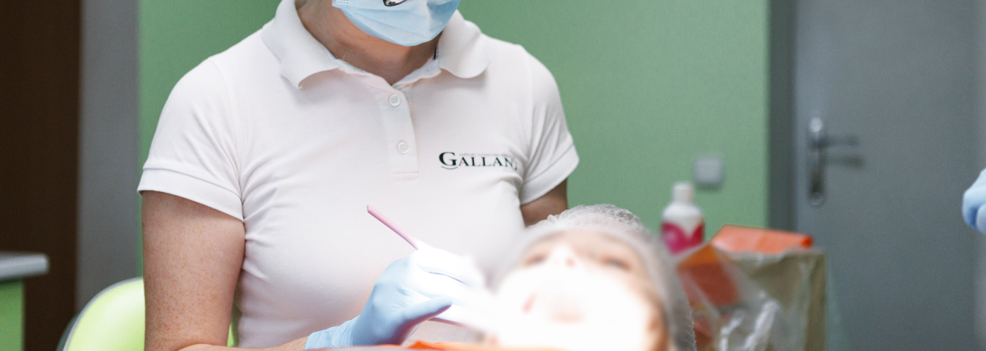
Root canal treatment under a microscope
In modern dentistry, performing root canal treatment under a microscope is a necessity. The microscope allows the dentist to control every step with maximum precision, ensuring accurate diagnosis and treatment. As a result, procedures are performed with higher quality and long-lasting outcomes. During treatment, the dentist removes old fillings, cleans the root canals, and fully restores the tooth. Thanks to the microscope, even the smallest structures are visible, reducing the risk of errors, trauma, and missed issues.
When Is Root Canal Treatment Under a Microscope Necessary?
Dental microscopes are used in various fields, including therapy, endodontics, prosthetics, and aesthetic dentistry. With magnification up to 30–40 times, a microscope allows for early detection of tooth decay and microcracks, precise canal cleaning, accurate preparation for crowns or veneers, perfect fit of restorations.
A microscope is used for:
- Tooth diagnostics (hidden caries, cracks, defects invisible on X-ray)
- Root canal treatment and filling
- Root treatment
- Fissure sealing
- Tooth restoration
- Crown and veneer preparation
- Veneer and implant placement
- Removal of granulomas, cysts, old fillings, and broken instruments
It enables a thorough examination of each tooth, detecting even the tiniest problems. This allows for early intervention and selection of the most effective treatment approach. Often, a microscope makes it possible to save a tooth that would otherwise require extraction.
Endodontics Under a Microscope
Root canals are tiny structures that connect the tooth pulp to surrounding tissues and house nerves and blood vessels. In deep caries cases with pain, endodontic treatment involves nerve removal, cleaning, and sealing of the canals. While X-rays were traditionally used, microscopes now allow for even more precise treatment in many cases — sometimes without needing X-rays.
Identifying Root Canals with a Microscope
Front teeth usually have one canal, while molars may have 2 to 4. These canals are extremely narrow and often curved, with diameters as small as 1 mm so it is impossible to see without magnification.
The dental microscope offers up to 40x magnification, helping the dentist accurately locate and evaluate the number, position, and even depth of root canals. This is crucial for thorough cleaning and filling, ensuring no branches are missed and avoiding complications or re-treatment. It also helps seal the canal completely without overfilling beyond the apex.
Repeat treatment of Root Canals Under a Microscope
In some cases, retreatment of root canals may be necessary if the previous treatment was not performed with sufficient quality. This involves opening the canals again and performing a new filling. Patients often come to specialists with complaints of pain in previously treated teeth, gum inflammation, and other complications. These issues may arise when not all canals were properly cleaned or if some remained unfilled. In some instances, a fragment of a medical instrument can even be found inside the root canal.
Using a microscope, the dentist can thoroughly examine the canals, identify all the issues in detail, and eliminate them. This allows for precise retreatment and new canal filling. In some cases, complete treatment may require several appointments. If inflammation is present, special medications are placed inside the canal, and the tooth is sealed with a temporary filling, which is replaced with a permanent one after a few days.
Canal Filling with Microscopic Precision
Ultra-fine instruments (0.6 mm or even 0.1 mm in diameter) are used during root canal treatment. Without a microscope, a dentist must rely on experience to choose tools. But with magnification, the entire process becomes visible and precise, allowing each canal to be sealed thoroughly and in stages.
Step-by-Step: How Root Canal Treatment Under a Microscope Works
- Diagnosis: The dentist examines mouth and may order an X-ray or CT scan. This helps assess the condition of the tooth and canals, detect inflammation, and plan treatment, especially useful for pulpitis, periodontitis, or to verify treatment results.
- Treatment Planning: Based on the diagnosis, an individual treatment plan is created.
- Preparation: Local anaesthesia is applied. A rubber dam is used to isolate the tooth, protecting the oral cavity from saliva and disinfectant solutions.
- Treatment Process: Under a microscope, the dentist cleans the canals, removes damaged tissue, measures the length and number of canals, performs mechanical and medical cleaning, then fills them with specialised materials for complete sealing and inflammation prevention.
- Treatment Control: X-rays or CT scans confirm correct sealing.
- Tooth Restoration: The final stage involves restoring the tooth’s shape and function with a filling, inlay, or crown — depending on the case.
If needed, a temporary filling and medication may be placed between visits. The number of appointments and treatment duration is always individual.
How long does root canal treatment under a microscope take?
The duration of root canal treatment under a microscope depends on the number and complexity of the canals in the tooth. Usually, the treatment takes 1–2 hours. Particularly complex cases may require 2–3 visits. The exact duration of the treatment can only be predicted by the dentist after a thorough examination of the condition of the teeth and oral cavity. It is advisable to perform a computed tomography (CT scan) of all teeth before root canal treatment under a microscope.

What is the purpose of a microscope in dentistry?
A microscope in dentistry is needed for treatment with magnification and additional lighting of the tooth canals. Multiple magnification allows detecting problems at early stages, which makes their resolution easier.
What are the indications for root canal treatment under a microscope?
- Acute or chronic pulpitis;
- Periodontitis;
- Infectious lesions of the root canals;
- Complex and advanced cases that require high precision treatment.
What are the stages of root canal treatment?
- Dentist consultation;
- Diagnostics;
- Anesthesia;
- Cleaning of the canals;
- Filling of the canals;
- Tooth restoration.
Testimonials
Dr. Vasyl Kulias is an excellent doctor at the Gallant Clinic. The treatment was carried out at the highest level - everything was professional and as painless as possible. I received much better care than I did abroad. I can even say that I came specifically to see this specialist. I confidently recommend him and am really grateful!